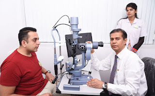
Dry Eye Treatment
Dry eye syndrome or Keratoconjunctivitis sicca (KCS), is a frequently encountered ocular problem. Dry eye syndrome is a clinical condition characterized by deficient tear production or excessive tear evaporation resulting in ocular discomfort. In addition to the term dry eye, which is now established worldwide, the specific term “ocular surface and tear disorder” has been suggested by Tseng and Tsubota.
High Risk Population for Dry Eye Disease
- Advanced age and female gender.
- Inflammatory disease (vascular, allergy, asthma)
- Autoimmune diseases (RA, Lupus, colitis)
- Peri and post menopausal women and Hormone Replacement Therapy patients.
- Diabetes Mellitus.
- Thyroid disease.
- Corneal transplantation
- Previous keratitis or corneal scarring
- Extra capsular or intracapsular large- incision cataract surgery.
- Laser in situ keratomileusis (LASIK) Systemic medications (diuretics, antihistamines, psychotropics, cholesterol-lowering agents)
- Contact lens wear
- Environmental conditions (allergens, cigarette smoke, wind, dry climate, air travel chemicals, some perfumes pollution).
Clinical Picture:
Symptom assessment plays a key role in dry eye diagnosis In addition, the symptoms agree poorly with the clinical tests and signs.
Patients have symptoms like burning sensation, irritation, itching, redness, blurred vision, photophobia and dryness. They are rated on a 5 point severity scale:
0 — no discomfort, 1 — trace, 2 — mild, 3 — moderate, 4 — severe
1. Tear film meniscus height — the marginal tear strip shows a full concave surface.
2. Tear film meniscus floaters — are tiny bits of debris, consisting of either dead epithelial cells or small fibrils of lipid contaminated mucin, being carried along the upper and lower lid meniscus.
3. Mucous Strands – are strings of lipid- contaminated mucin that have been pushed into the cul-de-sac by the shearing action of the lids.
4. Filaments — corneal filaments are checked with slit lamp.
5. Tear film breakup time (TBUT) is one of the commonest objective tests, utilized to make diagnosis of dry eye. After instillation of fluoresce in the tear film is observed with use of a cobalt blue filtered light of slit lamp and time that elapsed between last blink and appearance of 1“ break in the tear film is recorded. A result of <10 seconds suggest dry eye. Non-invasive tear break up time (NITBUT) methods have been developed using instruments like — keratometer, hand held keratoscope or tear scope.
6. Schirmer’s Test I — measures the total tear secretion (basic + reflex). Schirmer’s strip is made from What man’s no. 41 paper and measures 35x5mm. There is a bent at 5mm mark which goes into the cul-de-sac, at the junction of the middle and lateral 1/3 of the lower lid. After the patient has been described the procedure, gently blot the fornix and place the test strip at the proposed site. Patient is made to sit in a dimly light room and allowed to blink normally. The amount of wetting is measured after 5 min. Interpretation :
If wetting is >10mm = Normal.
If wetting is 10mm = Mild dry eye.
If wetting is 5-10mm = Moderate dry eye.
If wetting is 3-Smm = Severe dry eye.
If wetting is < 3 mm = Very Severe dry eye.
If wetting is 10mm = Mild dry eye.
If wetting is 5-10mm = Moderate dry eye.
If wetting is 3-Smm = Severe dry eye.
If wetting is < 3 mm = Very Severe dry eye.
7. Schirmer’s Test II — is used to ascertain reflex secretion. The procedure is similar to basal secretion test, but after the strips are installed, the unanaesthetised nasal mucosa is irritated by rubbing with dry cotton tipped applicator. The amount of wetting is measured after 2 minutes. Less than 15mm of wetting indicates failure of reflex secretion.
8. Rose Bengal staining – Rose Bengal is a vital dye and stains dying and desiccated epithelial cells, not protected by mucin layer.
Early or mild cases of KCS are detected more easily with Rose Bengal than fluoresce in staining and the conjunctiva is usually stained more intensely than cornea. Interpalpebral staining of the nasal and/or inferior paracentral cornea is seen in KCS, whereas a linear pattern of inferior conjunctiva and corneal staining is characteristics of MGD. Rose Bengal strip is inserted to inferior cul-de-sac. Patient is asked to blink several times. After 1 min, conjunctiva and cornea are examined using red free filter of slit lamp. For the purpose of evaluation eye 1S Divided into 3 zones- nasal conjunctiva, temporal conjunctiva and cornea. Amount of stain is recorded on the scale of 0 to 3, for a total possible score of 9 for each eye. Score of 3 or higher are consistent with KCS.
0- Negative
1- Scattered, minute staining.
2- Moderate spotty staining.
3- Diffuse blotchy staining.
1- Scattered, minute staining.
2- Moderate spotty staining.
3- Diffuse blotchy staining.
9. Lissamine green staining
10. Conjunctival impression cytology (CIC) Impression cytology is safe, non-invasive, tolerable and repeatable investigation in the diagnosis and management of dry eye disease.
Impression cytology of normal human bulbar conjunctival cells reveals relatively homogenous smaller cells with surface areas ranging from just 25-25.1m with an average of just over 100 um.
KCS shows following changes:
Squamous metaplasia
Stage 0 – Abundant goblet cell and mucin spots, small epithelial cells;
Stage 1 — Fewer goblet cells and mucin spots, small epithelial cells;
Stage 2 — Loss of goblet cells and mucin spots, enlarging epithelial cells;
Stage 3 — Enlarging or separating epithelial cells;
Stage 4 — Large separate epithelial cells with scattered keratinisation, pyknotic nuclei; Stage 5 — Large keratinized epithelial cells with pyknotic nuclei or loss of nuclei.
Loss of goblet cells.
Replacement of the normal corneal epithelial phenotype with an involved conjunctival epithelium.
Categories:


Comments
Post a Comment